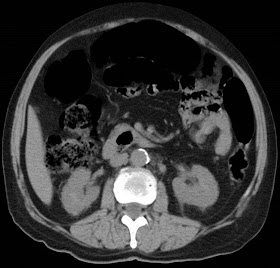
64 yrs female with painless hard lump in left breast with nipple retraction
64 yrs female with painless hard lump in left breast with nipple retraction
Signs of malignancy on mammogram are divided into major signs (conventional signs) and supporting signs (indirect signs).
Major Signs:
1. Spiculated Margins:
· Spiculated margins are a true diagnostic feature of malignancy.
· Strands of tissue are seen radiating out from an ill-defined mass, producing a stellate appearance.
· This appearance is pathognomonic of breast cancer.
· Spiculation represent retraction of tissue strands towards the tumor due to fibrosis - as a result of desmoplastic response.
· Sometimes, only the spiculation are seen without a mass
2. Clustered Microcalcifications:
· Mammography is the only technique capable of detecting microcalcification Microcalcification, even when found in isolation herald the presence of early stage breast cancer.
· Five or more calcifications, measuring less than 1mm, in a volume of one cubic centimeter, define a 'cluster'.
· The possibility of malignancy increases as the size of individual calcification decreases, the total number of calcification per limit area increases.
· It is the distribution and morphology of the calcifications, which defines their significance.
Supporting Signs of Malignancy
These indirect signs, though non-specific, signify enough risk to warrant intervention.
1. Poorly Defined Mass
· Most breast cancers are seen as poorly defined masses, without any mammographic features more suggestive of malignancy.
· Circumscribed masses with margins that are mostly well defined with only a portion ill-defined, are also managed as other ill-defined masses.
· There are a sizable number of benign breast masses whose margin appears to be poorly defined, and therefore are difficult to differentiate from malignancy resulting in the need to biopsy in order to detect early cancer.
2. Microlobulation
· Lobulations are usually associated with fibroadenomas.
· Increased number of lobulations, measuring few millimeters should be suspected for malignancy.
3. Architectural Distortion
· Breast cancer does not always produce a mammographically visible mass.
· Sometimes it produces just a localized cicatrization.
· If previous surgery and trauma to the breast can be excluded, there is high likelihood that the distortion is because of malignancy.
· Invasive carcinoma distorts the interface between breast and normal parenchyma due to desmoplastic response of host tissue to the malignancy.
4. Asymmetric Density
· Asymmetric density is the three dimensional area in which the density is greatest at the centre and fades towards the periphery trying to form a mass.
· In this situation, it is helpful to to view the mammograms of both breasts side by side.
5. Nipple Retraction
· Nipple retraction "over a short period of time" is suspicious of an underlying cancer.
6. Enlarged Axillary Lymph nodes
· Demonstration of large nodes is non-specific sign of malignancy.
· Involvement of the nodes(s) indicates worsening of prognosis.
BI-RADS Category
|
Assessment
|
Clinical Management Recommendation(s)
| |||
0
|
Assessment incomplete
|
Need to review prior studies and/or complete additional imaging
| |||
1
|
Negative
|
Continue routine screening
| |||
2
|
Benign finding
|
Continue routine screening
| |||
3
|
Probably benign finding
|
Short-term follow-up mammogram at 6 months, then every 6 to 12 months for 1 to 2 years
| |||
4
|
Suspicious abnormality
|
Perform biopsy, preferably needle biopsy
| |||
5
|
Highly suspicious of malignancy; appropriate action should be taken.
|
Biopsy and treatment, as necessary.
| |||
6
|
Known biopsy-proven malignancy, treatment pending
|
Assure that treatment is completed
|


