6 months old child with history of seizures. No history of hypoxic injury.
 |
| Axial T1 wt images show homogenous hypointensity in bilateral caudate nuclei and anteromedial thalami. |
 |
| Axial T2 wt images show homogenous hyperintensity in bilateral caudate nuclei, lentiform nuclei, antero medial thalami, cerebral peduncles and periaqueductal gray matter. |
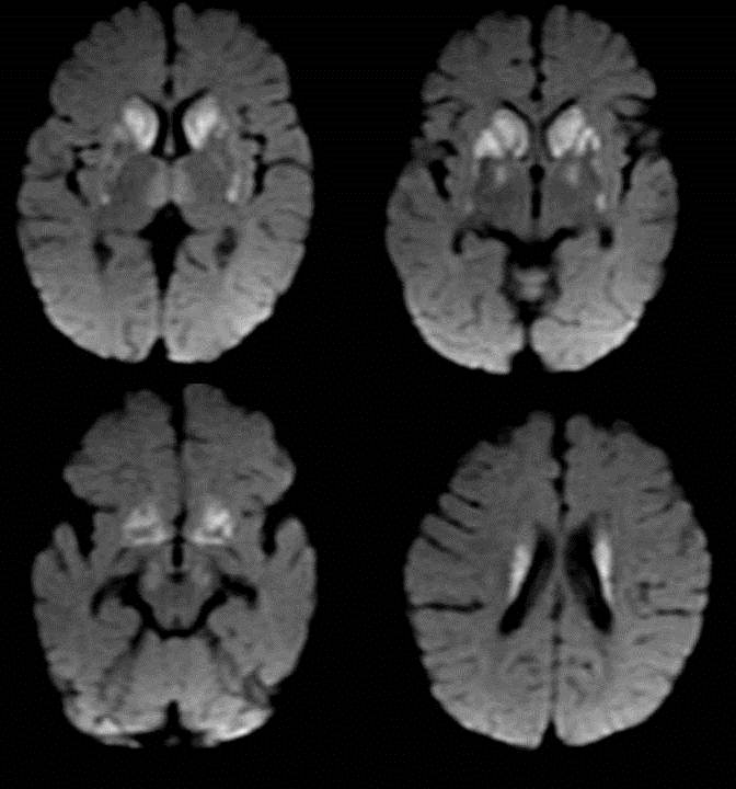 |
| All the above mentioned areas show restricted diffusion |
·
Leigh disease (subacute necrotizing encephalomyelopathy) is a
mitochondrial disease that results from a disorder in the respiratory chain
production of adenosine triphosphate.
·
Inherited, progressive, neurodegenerative disease of infancy or
early childhood with variable course and prognosis.
·
Clinical manifestations can be highly variable, affecting children
and (rarely) young adults and typically causing central hypotonia,
developmental regression or arrest, ophthalmoplegia, respiratory and bulbar
dysfunction, and ataxia.
·
Characteristic pathologic abnormalities include microcystic
cavitation, vascular proliferation, neuronal loss, and demyelination of the
midbrain, basal ganglia, and cerebellar dentate nuclei and, occasionally, of
the cerebral white matter.
·
MR imaging findings include symmetric areas of T2 prolongation in
the basal ganglia, periaqueductal region, and cerebral peduncles, with
putaminal involvement being a consistent feature.
·
The cerebral white matter is rarely affected. Enhancement may be
seen at MR imaging and may correspond to the onset of acute necrosis
·
When Leigh disease is suspected, MR spectroscopy (best performed
with long echo times) may reveal the presence of abnormally high lactate levels
in the basal ganglia, which together with elevated serum and CSF lactate levels
supports the diagnosis.
References
:
1. Leukodystrophy
in Children: A Pictorial Review of MR Imaging Features, May 2002 RadioGraphics, 22, 461-476
2. Differential
Diagnosis for Bilateral Abnormalities of the Basal Ganglia and Thalamus, January
2011 RadioGraphics, 31, 5-30
No comments:
Post a Comment
Please leave your comments