 |
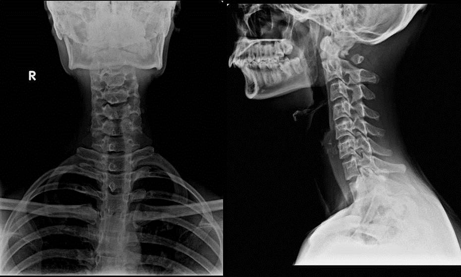
| AP and Lateral Radiographs of cervical spine showing absent posterior arch of atlas behind the lateral masses with free floating posterior tubercle. The lateral masses are well seen in AP radiograph. |
Embryology of atlas
· It has three primary ossification centers in the embryological period - an anterior center for formation of the anterior tubercle and two lateral centers from which the lateral masses and posterior arch form.
· In 7th week of gestation, the lateral centers extend dorsally to form the posterior arch.
· An additional center may be present posteriorly in the midline, forming the posterior tubercle of the C1 in second year of life in about 2% of the population.
· During maturation, the anterior arch of the C1 derives from usually one midline, but occasionally two or more ossification centers are seen radiologically in the first year of life.
· Ossification of the posterior arch usually proceeds perichondrally from two centers in the lateral masses, towards the midline ,and fusion occurs between the 3rd and 5th years of life.
· Incomplete fusion of the two hemiarches may be normal in the first 5-10 years. However, incomplete fusion may persist in 3 to 5% of patients.
· The anterior center usually fuses with the two lateral centers between 5 and 9 years age.
· In rare cases, a separate ossification center forms in the midline and fuses secondarily with the lateral masses forming the posterior arch.
· A posterior midline ossified tubercle develops if this center fails to fuse with the hemiarches.
· At least two different anomalies can develop during the ossification:
1) median clefts of the posterior arch of C1
2) varying degrees of posterior arch dysplasia
Currarino’s Morphologic Classification for C1 Hypoplasia

A Failure of posterior midline fusion of the two hemiarches
B Unilateral defect
C Bilateral defects
D Absence of posterior arch, with persistent posterior tubercle
E Absence of the entire arch, including the tubercle
Reference : Congenital Hypoplasia of the Posterior Arch of the Atlas, Turkish Neurosurgery 2011, Vol: 21, No: 1, 97-103


