 |
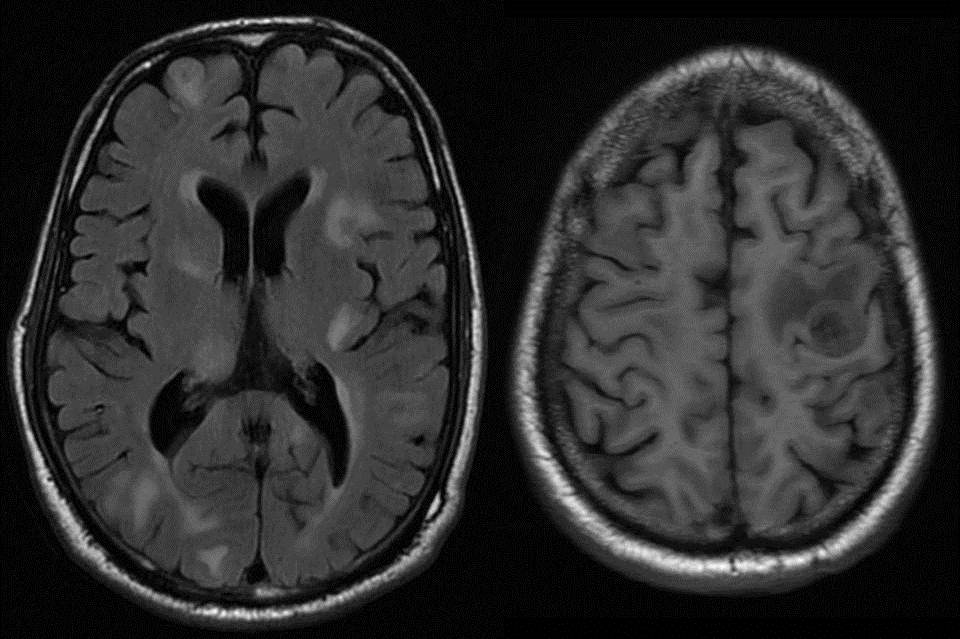
| Axial T2 FLAIR and T1 Wt images show multiple thick walled isointense lesions with central necrosis and surrounding edema, predominantly distributed at the corticomedullary junction. |
 |
| Axial contrast enhanced T1 Wt images show ring enhancement of all the lesions, with central dot like enhancement in some of them and irregular solid enhancement in the rest. Note the lesion in left sylvian fissure-s/o meningeal deposit. |
 |
| Axial and Coronal CT of chest revealed large irregular mass lesion in left perihilar region extending into apicoanterior segment with multiple metastatic nodules in the periphery of both lungs- s/o carcinoma lung with metastasis. |
·
Primary and metastatic tumors, on imaging, often manifest as
rounded, well-circumscribed, ring-enhancing lesions of variable sizes
surrounded by a variable amount of perifocal vasogenic edema.
·
Metastatic tumors are the most common intracranial neoplasm in
adults.
·
Lung cancer, breast cancer and melanoma account for the majority
of patients with metastasis in the brain.
·
The incidence of brain metastases has recently increased because
of several factors, including improved survival, better treatment of systemic
diseases and improved intracranial imaging techniques.
·
There were no characteristic computed tomography patterns for
specific systemic carcinomas, but epidermoid carcinoma frequently appeared as a
low-density lesion with a thin peripheral enhancing rim, and adenocarcinoma
appeared as a dense, homogeneous, round, enhancing nodule.
·
After treatment, focal cerebral parenchymal enhancement was the
most reliable sign of residual or recurrent tumor.
·
Metastatic lesions are typically subcortical, occurring in or near
the gray matter-white matter junction, and are usually associated with severe
perilesional edema.
·
MRI typically reveals mild T1 hypointensity with T2 hyperintensity
and fluid-attenuated inversion recovery hyperintensity at the site of the
lesion.
·
After contrast administration, a nodular ring pattern of
enhancement is seen.
·
Metastases from malignant melanoma may demonstrate T1
hyperintensity because of hemorrhagic or melanin components of the lesion.
·
Rapidly growing primary brain tumors, such as glioblastoma
multiforme or anaplastic astrocytoma, can present with many of the same imaging
characteristics as seen in metastatic lesions of the brain.
·
Most of the primary tumors are large in size and are often located
deep in the white matter.
·
Primary brain tumors frequently cross the midline. For example,
glioblastoma multiforme frequently crosses the midline by infiltrating the
white matter tracts of the corpus callosum.
·
Differential diagnosis of multiple ring enhancing lesions of
brain:

Reference :
Garg RK, Sinha MK. Multiple ring-enhancing
lesions of the brain. J Postgrad Med 2010;56:307-16
view similar cases:http://radfacts.blogspot.in/2012/06/neurocysticercosis.html
No comments:
Post a Comment
Please leave your comments