Friday 27 July 2012

ANTERIOR ABDOMINAL WALL INCISIONAL HERNIA

Lateral topogram of abdomen revealed herniation of bowel loop through anterior abdominal wall in infraumbilical region. Dilated jejunal loops with multiple airfluid levels.

TYPES OF ABDOMINAL HERNIAS:
1.     Groin hernias
a.      Inguinal
b.     Femoral
2.     Ventral hernias
a.      Umbilical
b.     Paraumbilical
c.      Epigastric
d.     Hypogastric
3.     Lumbar hernias
a.      Superior (Grynflett-Lesshaft)
b.     Inferior (petit)
4.     Incisional
5.     Others
a.      Interparietal
b.     Richter
c.      Litter
d.     Sciatic
e.      Obturator
f.       Perineal
Reference : Abdominal Wall Hernias: Imaging Features, Complications, and Diagnostic Pitfalls at Multi–Detector Row CT, November 2005 RadioGraphics, 25, 1501-1520

CEREBRAL METASTASIS- MULTIPLE RING ENHANCING LESIONS

Axial T2 FLAIR and T1 Wt images show multiple thick walled isointense lesions with central necrosis and surrounding edema, predominantly distributed at the corticomedullary junction.

Axial contrast enhanced T1 Wt images show ring enhancement of all the lesions, with central dot like enhancement in some of them and irregular solid enhancement in the rest. Note the lesion in left sylvian fissure-s/o meningeal deposit.

Axial and Coronal CT of chest revealed large irregular mass lesion in left perihilar region extending into apicoanterior segment with multiple metastatic nodules in the periphery of both lungs- s/o carcinoma lung with metastasis.

·        Primary and metastatic tumors, on imaging, often manifest as rounded, well-circumscribed, ring-enhancing lesions of variable sizes surrounded by a variable amount of perifocal vasogenic edema.

·        Metastatic tumors are the most common intracranial neoplasm in adults.

·        Lung cancer, breast cancer and melanoma account for the majority of patients with metastasis in the brain.

·        The incidence of brain metastases has recently increased because of several factors, including improved survival, better treatment of systemic diseases and improved intracranial imaging techniques.

·        There were no characteristic computed tomography patterns for specific systemic carcinomas, but epidermoid carcinoma frequently appeared as a low-density lesion with a thin peripheral enhancing rim, and adenocarcinoma appeared as a dense, homogeneous, round, enhancing nodule.

·        After treatment, focal cerebral parenchymal enhancement was the most reliable sign of residual or recurrent tumor.

·        Metastatic lesions are typically subcortical, occurring in or near the gray matter-white matter junction, and are usually associated with severe perilesional edema.

·        MRI typically reveals mild T1 hypointensity with T2 hyperintensity and fluid-attenuated inversion recovery hyperintensity at the site of the lesion.

·        After contrast administration, a nodular ring pattern of enhancement is seen.

·        Metastases from malignant melanoma may demonstrate T1 hyperintensity because of hemorrhagic or melanin components of the lesion.

·        Rapidly growing primary brain tumors, such as glioblastoma multiforme or anaplastic astrocytoma, can present with many of the same imaging characteristics as seen in metastatic lesions of the brain.

·        Most of the primary tumors are large in size and are often located deep in the white matter.

·        Primary brain tumors frequently cross the midline. For example, glioblastoma multiforme frequently crosses the midline by infiltrating the white matter tracts of the corpus callosum.

·        Differential diagnosis of multiple ring enhancing lesions of brain:


      Reference : Garg RK, Sinha MK. Multiple ring-enhancing lesions of the brain. J Postgrad Med 2010;56:307-16
view similar cases:
http://radfacts.blogspot.in/2012/06/neurocysticercosis.html

Thursday 26 July 2012

BENNETT'S fracture

Frontal radiograph of left wrist shows fracture base of 1st metacarpal with extension into articular surface. The fracture fragment maintains articulation with trapezium, rest of the metacarpal shows mild displacement laterally.


Rolando Fracture
  • Comminuted
  • Intra-articular
  • Fracture through base of thumb
  • Prognosis: worse than Bennett's fracture
    (difficult to reduce)

Bennett's Fracture
  • Intra-articular fracture/dislocation of base of 1st metacarpal
  • Small fragment of 1st metacarpal continues to articulate with trapezium
  • Lateral retraction of 1st metacarpal shaft by abductor pollicis longus

Monday 16 July 2012

BOUTENNIERE DEFORMITY


40 yrs female with inability to extend 3rd finger of left hand following trauma.


Frontal and lateral projections of 3rd finger of left hand showed flexion deformity of proximal interphalangeal joint and extension deformity of distal interphalangeal joint. moderate soft tissue swelling around the proximal phalanx. No evidence of fracture/ avulsion fragment.
High resolution sonography of 3rd finger of left hand revealed rupture of central slip of extensor digitorum tendon at its insersion onto the middle phalanx (1st image. see normal central slip of other finger for comparision). The two lateral slips are intact(2nd image). Axial images of normal and abnormal fingers showing edema at the site of disruption with absent tendon fibres and widely seperated lateral slips(3rd image).
Anatomy:
·        The extensor tendons to the four fingers pass across their respective metacarpophalangeal joints.
·        The deeper fibres of the tendons form the posterior capsule of the joints.
·        The bulk of each tendon passes freely across each joint. 
·        The tendons then broaden out and divide into three slips on the dorsal aspect of each proximal phalanx.
·        The central slip passes on to the base of the middle phalanx. 
·        The lateral slips diverge around the central slip and receive strong attachments from the interossei and lumbricals forming the extensor expansion.


Boutonnière (“button hole”) deformity:
·        Injury to the central tendon results in an imbalance in the extensor mechanism. 
·        Flexor digitorum superficialis is unopposed and it flexes the proximal interphalangeal joint.
·        This forces the head of the proximal phalanx between the lateral bands.
·        With time, the triangular ligament ruptures causing the lateral bands to displace volar to the axis of motion of the proximal interphalangeal joint and become flexors of the joint. 
·        The extensor hood retracts proximally, causing extension at the metacarpophalangeal and distal interphalangeal joints.
·        This results in  flexion of the proximal interphalangeal joint and hyperextension of the distal interphalangeal and metacarpophalangeal joints, known as the boutonnière (“button hole”) deformity.

·        Types of injury:
1.     Avulsion fracture of middle phalanx
2.     Partial rupture with stretching of central slip
3.     Complete rupture with seperation

Reference : Ultrasound imaging of finger tendons at the bedside in the emergency department, Southern Cross University ePublications@SCU.

Wednesday 11 July 2012

MIDGUT MALROTATION OF ADULT


Axial CECT of abdomen showing altered relation of SMA and SMV. The duodenal flexure is seen on the right of midline(not shown here). 


volume rendered images of bowel showed transverse orientation of ascending colon with caecum and appendix in right upper quadrant.

VR images of major vessels showed SMA to the right of SMV.

·        Intestinal malrotation can be broadly defined as any deviation from the normal 270° counterclockwise rotation of the midgut during embryologic development.
·        Malrotation results not only in the malposition of the bowel but also in the malfixation of the mesentery.
·        The normally broad mesenteric attachment is shortened to a narrow pedicle that predisposes the patient to the complication of midgut volvulus.
·        Internal hernia related to abnormal peritoneal fibrous bands (of Ladd) that attach to the right colon is another complication of malrotation seen in adults.
·        Conventional radiography is neither sensitive nor specific for malrotation, although right-sided jejunal markings and the absence of a stool-filled colon in the right lower quadrant may be suggestive of this finding.
·        The upper gastrointestinal barium series remains accurate for detection, and the rules familiar to pediatric radiology also apply for adults—that is, the duodenal—jejunal junction fails to cross the midline and lies below the level of the duodenal bulb.
·        An abnormal junction in an adult should not be dismissed as a normal variant.
·        Contrast enema examination usually shows malposition of the right colon, but the cecum may assume a normal location in up to 20% of patients.
·        The contrast enema findings are also nonspecific because cecal location can be variable without malrotation.
·        Many cases of quiescent malrotation in adults are currently being detected on cross-sectional imaging performed for various unrelated reasons.
·        CT not only shows the intestinal malpositioning seen on barium studies but also depicts associated extraintestinal findings not evident on conventional examinations. For example, deviation from the normal relationship between the SMA and SMV is a useful indicator of malrotation.
·        In most patients with quiescent malrotation, the SMA and SMV will assume a vertical relationship or show left—right inversion.
·        Analogous findings can be seen on sonography.
·        Abnormalities of SMA—SMV orientation are not entirely diagnostic, however, because some patients with malrotation will have a normal relationship, and a vertical or inverted relationship can also be seen in patients without malrotation.
·        Therefore, isolated detection of such an abnormality is not sufficient for diagnosis but should warrant closer examination of the bowel.
·        Finally, inspection of the pancreas in malrotation will reveal underdevelopment or absence of the uncinate process.
·        Acute complications of malrotation:
1.     Midgut volvulus
2.     Internal hernia
·        Regardless of patient age, surgical treatment of quiescent malrotation should be considered because surgery remains the only real safeguard against complications.
Reference : Intestinal Malrotation in Adolescents and Adults: Spectrum of Clinical and Imaging Features, AJR December 2002 vol. 179 no. 6 1429-1435.


METACHROMATIC LEUKODYSTROPHY

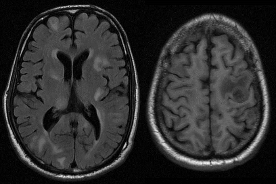
Axial T2 FLAIR images showing diffuse hypointensity of bilateral frontoparietal white matter with sparing of subcortical U fibres. There is sparing of perivascular white matter giving a "TIGROID" or "LEOPARD SKIN" appearance

Axial and Sagittal T2 Wt images show extent of hyperintense signal in bilateral fronto temporo parieto occipital white matter from deep periventricular region to subcortical region
with sparing of U fibres.

Axial DWI and sagittal T1 Wt images: Mild restricted diffusion noted along the advancing edge of dysmyelination. Note the involvement of distal body and splenium of corpus callosum
in the disease process on T1 Wt image.

Classification of Leukodystrophies:


·        Metachromatic leukodystrophy is an autosomal recessive disorder caused by a deficiency of the lysosomal enzyme arylsulfatase A.
·        This enzyme is necessary for formation of myelin sheath.
·        In metachromatic leukodystrophy, sulfatides accumulate in various tissues, including the brain, peripheral nerves, kidneys, liver, and gallbladder.
·        The accumulation of sulfatides within glial cells and neurons causes the characteristic metachromatic reaction.
·        Metachromatic leukodystrophy is diagnosed biochemically on the basis of an abnormally low level of arylsulfatase A in peripheral blood leukocytes and in urine.
·        Three different types of metachromatic leukodystrophy are recognized according to patient age at onset:
1.     Late infantile
2.     Juvenile
3.     Adult
·        The most common type is late infantile metachromatic leukodystrophy, which usually manifests in children between 12 and 18 months of age and is characterized by motor signs of peripheral neuropathy followed by deterioration in intellect, speech, and coordination.
·        Within 2 years of onset, gait disturbance, quadriplegia, blindness, and decerebrate posturing may be seen.
·        Disease progression is inexorable, and death occurs 6 months to 4 years after onset of symptoms.
·        At T2-weighted MR imaging, metachromatic leukodystrophy manifests as symmetric confluent areas of high signal intensity in the periventricular white matter with sparing of the subcortical U fibers.
·        No enhancement is evident at computed tomography or MR imaging.
·        The tigroid and “leopard skin” patterns of demyelination, which suggest sparing of the perivascular white matter, can be seen in the periventricular white matter and centrum semiovale.
·        The corpus callosum, internal capsule, and corticospinal tracts are also frequently involved.
·        The cerebellar white matter may appear hyperintense at T2-weighted MR imaging.
·        In the later stage of metachromatic leukodystrophy, corticosubcortical atrophy often occurs, particularly when the subcortical white matter is involved.
Reference : Leukodystrophy in Children: A Pictorial Review of MR Imaging Features, May 2002 RadioGraphics, 22, 461-476.

Tuesday 10 July 2012

SPINAL ARACHNOID CYST

Sagittal and Axial T2 wt images of lumbar spine showing a well defined oval fluid intensity extradural lesion with few internal septations in left lateral aspect of thecal sac from D12 to L2 level with extension into neural foraminae at D12-L1 and L1-2 levels.
Mild smooth displacement of thecal sac to right side.

According to the classification described by Nabors et al for spinal meningeal cysts, extramedullary cysts of the spinal canal can be divided into three main groups.

Group I : Meningeal cysts.

                  Classified into three types:

1.     Extradural meningeal cysts that contain no neural tissue (arachnoid cysts-Ia and sacral meningoceles-Ib)

2.     Extradural meningeal cysts that contain neural tissue (tarlovs cyst, cord herniation into cyst)

3.     Intradural meningeal cysts.

Group II : Nonmeningeal epidural cysts

(includes nonneoplastic lesions such as juxtaarticular synovial cysts, pigmented villonodular synovitis, and, rarely, herniated disks, as well as neoplastic lesions such as dermoids, cystic nerve sheath lesions, and metastases. In addition, trauma, hemorrhage, and inflammation can produce cystlike changes that are visible on cross-sectional imaging)

Group III : Neurenteric cysts.


Spinal Arachnoid cyst:

·        Probably arise from the herniation of the arachnoid through congenital or acquired dural defects.

·        The cysts begin as diverticula and subsequently enlarge, possibly because of a valvelike mechanism.

·        Postural changes and Valsalva's maneuver may produce symptoms.

·        These cysts usually arise in the mid-to-lower thoracic spine, often at the junction of the meninges and the dorsal nerve root sleeves proximal to the dorsal root ganglia.

·        Type 1A lesions most commonly project dorsally and may partially protrude into the adjacent neural foramen.

·        “Arachnoid cyst” is a histologic diagnosis, and the cyst has either no epithelial lining or an attenuated lining of arachnoidlike cells.

·        In the imaging literature, the terms “arachnoid cyst” and “meningeal cyst” are used interchangeably.

Reference : CT Myelography and MR Imaging of Extramedullary Cysts of the Spinal Canal in Adult and Pediatric Patients, AJR January 2002 vol. 178 no. 1 201-207